By Pekka Ruusuvuori and Alison Ollikainen
When it comes to modern technology, machine learning is a hot topic in the field of artificial intelligence and has already been used in image analysis for a long time. Rapidly advancing from the days of teaching a machine to recognize the difference between cats and dogs, Pekka Ruusuvuori is implementing this new tool to digital pathology applications. His focus is on cancer research, developing algorithms and computational models to find a faster approach to diagnostics.
Pekka Ruusuvuori is an Adjunct Professor, leader of the Bioimage informatics group and an affiliate of the Computational biology group at Tampere University. The group aims to uncover the molecular basis of human cancers and to generate new avenues for treatment and diagnostics by integrating both experimental and computational expertise within the group.
Here, Pekka tells us more about the benefits of machine learning in cancer research…
What does artificial intelligence and deep learning mean in the context of image analysis?
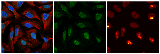
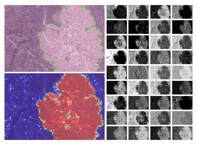
Machine learning has been used very intensively in image analysis for a long time already. It is an efficient approach for turning the information within pixels into decisions about the content of the images. For example, the question “is this a cancer cell or part of normal tissue?” could be answered with the help of machine learning model, given that the model has been trained for that purpose. Training is really the key ingredient here. Often, the hardest part is to get enough training material, for which we know the true class, like “cancer” or “normal”.
Recently, deep learning with neural networks has proved to be so accurate in many biomedical applications that it has reached superhuman accuracy. Whether to call it artificial intelligence or just machine learning, I guess it has something to do with what kind of audience is targeted. 😊
If we don’t know something exists within an image, is it possible for a machine to find something we haven’t taught it to look for?
While deep learning with, for example, convolutional neural networks, is indeed a powerful approach for classification, what they essentially do is they capture the characteristics of the image content in a feature space within the network. This means that the neural networks can be used for analyzing the image content even when the network would originally be trained for a completely different task. This kind of transfer learning with pre-trained networks works surprisingly well for many tasks and sometimes makes the training phase a lot easier.
How do you use AI & deep learning in your research?
Our Bioimage informatics group uses AI & deep learning for many tasks; breast and prostate cancer diagnosis and grading, detection of mutations in tumors, cross-staining modeling, single cell detection, cell phenotyping in leukemia, heart & vasculature diseases and for understanding human cells in general. One could say AI and machine learning are the key components in our research.
What is your hope for this approach to image analysis to be used in the future of medicine and research?
What we want to achieve is better understanding of what the images can tell about the cells and tissues. Imaging is one of the most important measurement techniques, and our group wants to get the most out of the images for the benefit of cancer researchers, pathologists, and basically for anyone doing quantitative microscopy of cells and tissues. Some of our solutions are already available in software used in routine clinical pathology.


One thought on “Is this cancer? Training Machines to Identify Cancer Cells from an Image”
Comments are closed.