By: Heli Kuisma
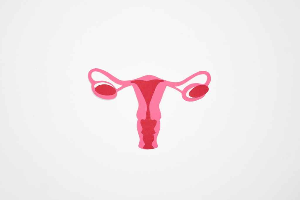
Fibroids, also known as uterine leiomyomas or as “uterine stones” (if you asked Hippocrates 460 BC) are extremely common benign tumors affecting approximately 70% of women by the age of 50. Although not life threatening, one in four of the patients have symptoms such as pain, abnormal menstrual bleeding, or infertility problems. Those with severe symptoms often need surgery as no curative drug-treatment exists. In fact, hysterectomy (the removal of the uterus) is often performed. The estimations for the yearly cost of treating fibroids in the USA range from 2 billion to 34 billion dollars. Considering the huge impact this tumor type has on both well-being and economy, these tumors remain severely understudied. A simple Google search for “fibroids” returns 59 million hits while a similar search for “prostate cancer” has almost 2 billion hits and “breast cancer” almost 4 billion hits.

At the forefront of fibroid research
Our research team, the Tumor Genomics Group led by Academy Professor Lauri Aaltonen from the University of Helsinki, has for a long time had a pioneering role in the study of fibroids. The cornerstone of this work has been a continuous collection of these tumors: after the surgery, fibroids from patients willing to participate in our research project are collected and stored for research purposes. Over the years, one fibroid sample can provide material for several analyses. The first fibroids in the Aaltonen group were collected already in 2001, and the work continues. To date, we have a collection of over 2000 fibroids from over 700 patients. We are extremely grateful for these patients because without their contribution our research would be impossible.
Similar on the surface level but different inside
During surgery most fibroids look similar: they are rubbery, bulging masses clearly separated from the surrounding normal uterine tissue. On the genetic level, however, fibroids are by no means identical and have different genetic defects as their root cause. Already in 1995, Schoenmakers and colleagues discovered aberrations affecting the HMGA2 gene in a subset of fibroids. In 2002 work by us and others led to the discovery of mutations in the FH gene. In 2011 we discovered mutations in the MED12 gene in a whopping 70% of fibroids. The exact mechanism of how these genetic alterations lead to the development of fibroids is, however, in many parts still beyond our current understanding.
Reaching into the unknown
Although HMGA2, FH, and MED12 alterations are the root cause in 90% of all fibroids, this still leaves 10% with no known cause. We were intrigued by these tumors that we called “the unknown fibroids”. This led us to study the gene expression changes in a large set of tumors including over one hundred of the unknown fibroids with RNA sequencing. RNA-sequencing gives information on two levels: on one hand, we get the quantity of the RNA corresponding to the expression level of the gene. On the other hand, we can decipher the mutations that were present in the DNA sequence which acted as the template for the RNA. By carefully combing the data of the unknown fibroid we saw a pattern emerging: there was a group of unknown tumors which seemed very similar to each other when looking at their gene expression. On mutation level all of these tumors did not seem to share a common mutation. Instead, several had mutations in genes such as YEATS4, DMAP1, and ZNHIT1. We understood that these genes encode for proteins that work together in the same protein complex – the SRCAP complex.
A brand new mechanism for tumor initiation
The SRCAP complex plays a key role in the regulation of histones. Histones are essential packaging proteins around which the DNA strand coils. This ensures the two meters of DNA per single cell really fits inside in an organized manner. In more detail, SRCAP complex works to add a specific type of histone, H2A.Z, into the DNA. After detecting the mutations we wanted to see if H2A.Z is somehow affected in these fibroids. We decided to study the levels of H2A.Z in our samples with immunohistochemistry (IHC). In IHC an antibody, a specific protein that can recognize other proteins, is used to detect the possible presence of the H2A.Z histones in a tissue sample. Indeed, we could not detect any or only very little H2A.Z in the samples with the SRCAP complex mutations – but plenty in normal uterine tissue – confirming that these newly discovered mutations have a biological effect on the respective fibroids.

Our results meant that we had discovered a brand new mechanism capable of causing tumors. More data was needed to understand this further and to compare the SRCAP mutated fibroids to other types of fibroids. The collected samples were utilized in a cutting edge laboratory work from which massive amounts of data were created for our data-analysis team. These included methylation data (to detect DNA methylation, a specific DNA alteration that can have a drastic effect on the function of the genes), ChIP-seq (to detect the DNA regions in which H2A.Z is located), and ATAC-seq (to discover DNA regions where the DNA is less tightly coiled around the histones). By integrating all the available data we discovered that the broken H2A.Z leads to deregulation of bivalent genes. Bivalent genes refer to a set of genes that are important during the development of an individual. They are typically in a paused state in our cells but with specific signals can be turned on very quickly and drive the development into, for example, a specific cell type. The situation is analogous to a paused video which can be quickly started with a press of a button. Interestingly, bivalent regions were not only highlighted in the SRCAP complex mutated fibroids but through different mechanisms also in other fibroids. The discovery of the SRCAP complex mutated fibroids as well as their biological properties was published in Nature in 2021.
Genomics research for the help of the patients
Understanding the differences between fibroids facilitates the development of drugs for these tumors: one treatment may not work for every fibroid. Deep knowledge of the genetics of fibroids can also help to find features that are shared by them all. We can also highlight features present only in one type of fibroid and search for targeted treatments specific to those features. Surgical treatment is never completely risk-free and requires time for recovery. In addition, hysterectomy is never a good option for women wanting to have children. An efficient drug for fibroids would thus be life-changing for millions of women, and we are vigorously working towards that goal.

Want to learn more about the Finnish Center of Excellence in Tumor Genetics?
Find the latest tweets, videos and blog posts for a peek into the everyday life in cancer research
by following us on Instagram (@tumorgenetics) and Twitter (@CoEinTG)
You can also subscribe to our YouTube channel!
