
By: Anni Kivinen and Aliisa Tiihonen
Cells are known to interact actively with their environment. Simple single-celled organisms must sense variations in their surroundings and react to them accordingly. Since the emergence of multicellular organisms, the importance of cell-cell signaling and interaction has become significant to assure that the organism responds to stimuli in the necessary way. For example, the function of our whole nervous system and immune system is completely dependent on the interaction of cells.
Spatial location matters.
The means of cell communication are diverse. Cells can receive signals from other cells over short and long distances. As some signaling is restricted to the immediate neighborhood of a cell, the spatial organization of cells in tissue determines their interaction and function. It is known that cancer for example alters the tissue structure and disturbs the cell communication.
Different cell types express different sets of genes and thus the physical location of cells can be studied by analyzing the expression of cell type specific marker genes. Recent developments have made spatial technologies even more efficient: instead of focusing on a handful of genes, it is now possible to study all the genes expressed in a given cell (the whole transcriptome) in their native location. The fact that Nature Methods named spatially resolved transcriptomics as Method of the Year 2020 illustrates the scientific community’s interest and expectations towards the technology.
Spatial methods add to the family of transcriptomics.
Previous single-cell transcriptomics methods have enabled researchers to study the gene expression of single cells instead of averaged expression of genes in a sample. These methods however contain a step where the tissue is dissociated, and thus spatial information of the cells is lost. A widely used allegory to describe different transcriptomic methods is that the data in bulk RNA sequencing is like a fruit smoothie, in single-cell RNA sequencing like a fruit salad, and in spatial transcriptomics like a fruit tart where each piece of fruit has its own distinct place with regard to other pieces of fruit.

What is there to discover?
Spatial transcriptomics can be used to study the tumor microenvironment, and a large number of studies have been published already in this area. It is important to understand how cancer alters the surrounding tissue in order to understand tumor progression and why some tumor cells are resistant to treatments. Spatial transcriptomics can also be used to study tumor subclones and help to find new cell type specific biomarkers. In addition to cancer research, spatial transcriptomics has been widely used to study tissue development and homeostasis. Just recently, spatial transcriptomics was used to study the effect and spreading of COVID-19 virus in lung tissue.
Different spatial technologies utilized at Tampere University.
We started experimenting with spatial transcriptomics at Tampere University with the 10X Genomics Visium method. Unlike the earlier methods, where the location of cells is studied by imaging the fluorescence of the selected marker genes in situ, Visium captures RNA molecules from the sample in situ and the library is then sequenced ex situ. Each RNA molecule can be traced back to its original location on the tissue sample based on spatial barcodes (pieces of sequence) that are incorporated in the RNA capture oligonucleotides.
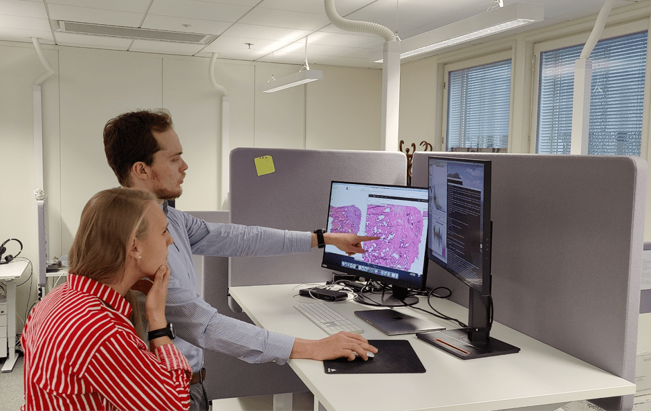
The technology was piloted in Nykter and Granberg Labs in October 2020 with prostate and brain cancer samples and we have expanded the analysis to additional samples after that. With this new technology, we have had to learn to use new tools both in the wet lab and on the computational side, so it has been an exciting journey for us.
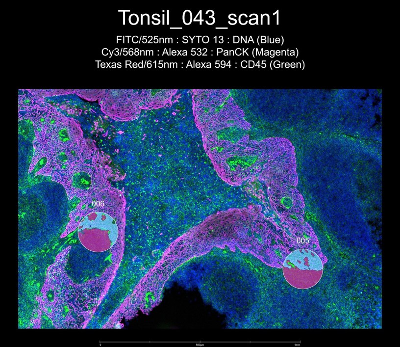
During the spring 2021, Tampere University acquired a GeoMx Digital Spatial Profiler, technology designed for spatial omics experiments from NanoString. GeoMx technology allows selecting specific tissue compartments or cell types for deeper analysis through fluorescent morphology staining. Ultraviolet light is then very precisely directed to each of the selected areas, resulting in the release of UV-cleavable oligomers, which are then collected for further analysis. The oligomers carry the molecular information needed to subsequently quantify the presence of different transcripts or proteins in the selected areas in the sample. The technology can be used for both fresh frozen and Formalin Fixed Paraffin Embedded (FFPE) samples, making it widely applicable for different research purposes.

In Tampere, GeoMx is planned to be used to study for example brain tumor micorenvironment. To this day, there is a lack of effective treatments for CNS (central nervous system) malignancies. Therefore, research on these tumors is highly needed in order to push the treatment development forward. Spatial transcriptomics will be used to especially analyze immunosuppression in the tumors, that not only blocks effective immune response against the tumor, but also supports the tumor growth. Spatial transcriptomics offers an excellent way to study the tumor and immune cell interactions and activity.
The future of spatial transcriptomics.
Spatial transcriptomics is a rapidly evolving field and new advances in the technologies and the analysis tools for spatial data are being developed as you read this post. If the technologies keep developing as expected, some day we might be able to go beyond the transcriptome, to spatially resolved proteome, epigenome, and metabolome to get a ‘multiomic’ spatial aspect. And perhaps in the future spatial transcriptomics could even help pathologists and oncologists to analyze tumorous areas in tissue samples for improved patient diagnostics and care.

Want to learn more about the Finnish Center of Excellence in Tumor Genetics?
Find the latest tweets, videos and blog posts for a peek into the everyday life in cancer research by following us on Instagram (@tumorgenetics) and Twitter (@CoEinTG)
You can also subscribe to our YouTube channel!

2 thoughts on “From Smoothies to Fruit Tarts: The Development of Spatial Transcriptomics.”
Comments are closed.